Sep
3D/4D, National Ultrasound
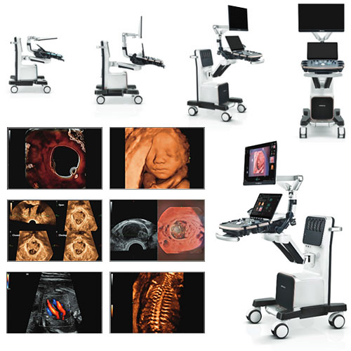
An Introduction to 4D Ultrasound: What Sets It Apart?
When it comes to medical imaging, ultrasound has been a game-changer, allowing healthcare professionals to peer inside the human body without the need for invasive procedures. Most of us are familiar with 2D and 3D ultrasounds, commonly used in prenatal care, but there’s another player in town – the 4D ultrasound. In this article, we’ll dive into the intriguing world of 4D ultrasound and explore what makes it different from its predecessors.
 The Basics of Ultrasound
Foundations of Ultrasound Imaging
Ultrasound imaging, or sonography, relies on the transmission of high-frequency sound waves into the body. These waves bounce back as echoes when they encounter different tissues, creating a real-time image on a screen.
2D Ultrasound
Traditional 2D ultrasound generates two-dimensional images, offering valuable insights into the structure and position of organs and, most notably, during prenatal scans.
3D Ultrasound
3D ultrasound takes it up a notch by adding depth to the images. This technology is especially valuable in obstetrics for assessing fetal development.
The Leap to 4D Ultrasound
What Exactly Is 4D Ultrasound?
4D ultrasound builds upon the foundation of 3D ultrasound but introduces an element that sets it apart – real-time movement. In simpler terms, it creates a 3D image that’s in motion, delivering a live-action view of the subject being scanned.
The Inner Workings
Much like its predecessors, 4D ultrasound employs sound waves to generate images. However, the advanced software and hardware used in 4D technology permit continuous scanning, resulting in a moving, time-lapsed image.
Real-Time Visualization
One of the standout features of 4D ultrasound is its ability to offer real-time visualization. Physicians and technicians can witness a baby in the womb moving, the beating of a heart, or the flow of blood through vessels as it unfolds.
Applications of 4D Ultrasound
Obstetrics and Prenatal Care
4D ultrasound is especially popular in obstetrics. Expectant parents can witness a detailed, real-time view of their developing baby, from facial expressions to tiny limb movements. It offers a more immersive and emotional experience during pregnancy.
Cardiology
In cardiology, 4D ultrasound plays a vital role in assessing heart function. It provides insights into the movement of heart valves, the efficiency of blood flow, and any structural irregularities.
Gynecology
Gynecologists rely on 4D ultrasound to examine the female reproductive system. It aids in diagnosing and monitoring conditions like fibroids, cysts, and uterine abnormalities.
Musculoskeletal Imaging
Beyond its use in pregnancy and cardiology, 4D ultrasound proves invaluable in musculoskeletal imaging. It helps orthopedic specialists visualize joint movement and assess muscle function in real-time, aiding in the diagnosis and treatment of injuries and conditions.
The Advantages of 4D Ultrasound
Enhanced Visualization
The real-time, moving images provided by 4D ultrasound offer a more comprehensive view of anatomy and function. This makes it easier to detect abnormalities and assess dynamic processes.
Improved Patient Understanding
Patients often find 4D ultrasound more engaging and easier to comprehend. Watching their baby’s movements or the beating of their heart can deepen their connection with the medical process.
Enhanced Diagnostic Accuracy
In many cases, 4D ultrasound can enhance diagnostic accuracy due to its ability to capture real-time changes and movements within the body.
Limitations and Considerations
While 4D ultrasound offers numerous advantages, it’s essential to acknowledge its limitations. It may not always be necessary for every medical imaging scenario and can be more expensive and time-consuming than traditional 2D ultrasound. Additionally, not all medical facilities may have 4D ultrasound equipment readily available.
4D ultrasound is a remarkable advancement in medical imaging technology. Its real-time, moving images provide valuable insights into the human body’s structure and function, making it a powerful tool in fields such as obstetrics, cardiology, gynecology, and musculoskeletal imaging. While it may not be required for every situation, its ability to offer enhanced visualization, improve patient understanding, and increase diagnostic accuracy makes it an exciting addition to the world of medical imaging. As technology continues to evolve, we can expect even more exciting developments in the field of ultrasound imaging.